Introduction
In this article, Dr Shree Rao talks about auricular seroma. She is the Best Doctor for Cochlear Implants.
Auricular seroma is a collection of serous fluid that accumulates between the layers of tissue in the ear, causing a swelling or lump on the auricle or pinna. The thin skin in this region makes it vulnerable to injuries, and trauma, infections, medical procedures, and underlying conditions can contribute to the development of auricular seroma. Throughout this article, readers will explore the causes, symptoms, diagnosis, and treatment options for auricular seroma, with a focus on how Dr. Shree Rao, an experienced ENT specialist, can assist in effectively managing and finding relief from this condition. Let them embark on this journey of understanding and take proactive steps towards ear health and well-being.
Symptoms of auricular seroma
Auricular seroma is characterized by specific signs and symptoms that can help identify its presence. Being aware of these indicators is crucial for early detection and timely treatment. Here are the common signs and symptoms associated with auricular seroma:
- Swelling and puffiness of the ear – One of the primary signs of auricular seroma is noticeable swelling and puffiness of the affected ear. The ear may appear larger than usual due to the accumulation of fluid between the layers of tissue.
- Fluid accumulation behind the ear – Auricular seroma often leads to the formation of a fluid-filled lump behind the ear. This accumulation of serous fluid creates a palpable mass that can be felt upon touching the area.
- Tender or painful sensation in the affected area – The presence of auricular seroma may cause tenderness or pain in the affected area. Patients may experience discomfort, particularly when touching or applying pressure to the swollen ear.
- Changes in ear shape or contour – As the serous fluid accumulates, the shape or contour of the ear may be altered. The affected ear may appear misshapen or have an irregular contour due to the swelling.
If individuals notice any of these signs and symptoms in their or their child’s ear, it is essential to seek medical attention promptly. Early intervention can prevent complications and promote a faster recovery, ensuring optimal ear health and well-being.
Diagnosing auricular seroma
Here are the key methods used in the diagnostic process:
The diagnostic process typically begins with a thorough physical examination of the affected ear. The medical professional will visually assess the ear for signs of swelling, puffiness, and fluid accumulation. They will also inquire about the patient’s medical history and any recent injuries or trauma to the ear.
To confirm the presence of fluid accumulation and assess the extent of the seroma, imaging tests such as ultrasound or magnetic resonance imaging (MRI) may be conducted. Ultrasound uses sound waves to create images of the ear’s internal structures, while MRI provides detailed cross-sectional images of the ear and surrounding tissues.
In some cases, the medical professional may perform an aspiration procedure to extract a sample of the serous fluid from the ear. This fluid sample is then sent for laboratory analysis to determine its composition and rule out other potential conditions.
A comprehensive and accurate diagnosis of auricular seroma enables the medical professional to devise an appropriate treatment plan tailored to the individual’s needs.
Causes and risk factors of auricular seroma
Auricular seroma can arise from various factors and conditions, each contributing to the development of fluid accumulation in the ear. Understanding these causes and risk factors is essential for identifying potential triggers and addressing them effectively. Here are the key factors associated with auricular seroma:
- Trauma or injury to the ear – One of the primary causes of auricular seroma is trauma or injury to the ear. Accidents, falls, or direct blows to the ear can lead to damage to the blood vessels and tissues, resulting in the accumulation of serous fluid in the affected area.
- Surgical complications after otoplasty or ear surgery – In some cases, auricular seroma may occur as a complication following otoplasty or other ear surgeries. Fluid accumulation can be a result of post-operative tissue inflammation and disrupted blood flow.
- Inflammatory conditions and infections – Inflammatory conditions of the ear, such as cellulitis, can contribute to the development of auricular seroma. Additionally, ear infections that are left untreated or inadequately managed may lead to fluid buildup in the ear.
- Hematomas and seromas – It’s important to differentiate between hematomas and seromas. While both involve fluid collection, hematomas consist of blood, whereas seromas contain serous fluid. Hematomas usually result from blood vessel injuries, while seromas are commonly associated with post-traumatic fluid accumulation.
Treating Auricular Seroma
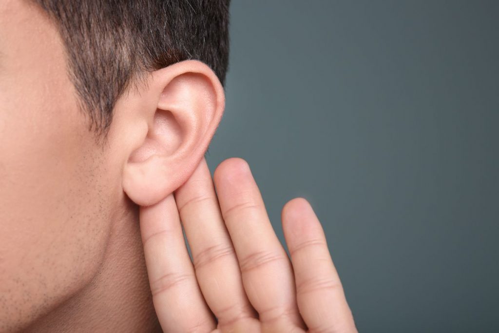
Here are the key medical and surgical options for treating auricular seroma:
- Aspiration – Aspiration is a common non-surgical approach for managing auricular seroma. In this procedure, a medical professional uses a needle and syringe to carefully drain the accumulated serous fluid from the affected area. Aspiration is generally a quick and minimally invasive procedure, providing immediate relief to the patient.
- Compression dressing – After aspiration, medical professionals may apply a compression dressing to the ear to encourage the absorption of any remaining serous fluid. The dressing applies gentle pressure to the area, helping to prevent fluid from re-accumulating and promoting the healing process.
- Surgical drainage – In some cases of auricular seroma, surgical drainage may be necessary, especially when the fluid accumulation is persistent or extensive.
Preventing recurrence is a crucial aspect of treating auricular seroma. Medical professionals work closely with patients to identify and address potential risk factors. Recommendations may include careful wound care after ear surgery, prompt management of infections or inflammation, and appropriate follow-up visits to monitor the healing process.
Tips for preventing auricular seroma
- Proper ear protection during physical activities – Individuals should wear protective headgear, such as helmets or headbands, while participating in contact sports or activities with a higher risk of ear injury, to minimize the risk of seroma formation.
- Adhering to post-operative care after ear surgery – Following the post-operative care instructions provided by the surgeon after ear surgery, such as otoplasty or reconstructive procedures, is essential. This includes proper wound care, avoiding unnecessary pressure on the ears, and attending follow-up appointments.
- Avoiding traumatic injuries to the ear – Being cautious and avoiding activities that may pose a risk to the ears can prevent accidental injuries that could lead to auricular seroma. Individuals should refrain from rough handling of the ears, pulling, or applying excessive pressure.
Conclusion
In conclusion, understanding auricular seroma is essential for early detection and prompt management of this condition. Dr. Shree Rao’s expertise and compassionate care can guide patients towards effective treatment and improved ear health. By recognizing the signs and symptoms, seeking timely medical attention, and following preventive measures, individuals can reduce the risk of auricular seroma and its recurrence. Dr. Shree Rao’s dedication to patient well-being and her comprehensive approach make her a trusted ally in the journey of ear health and overall well-being. Let Dr. Shree Rao’s expertise be your guiding light towards a healthier and happier life.

Why consult EarSurgeon, Dr. Shree Rao?
Dr. Shree Cuddapah Rao is acclaimed as one of the best pediatric ENT specialists in Hyderabad. With 10+ years of deep domain experience in the field of ENT, she is the director at Dr. Rao’s ENT Super Specialty Hospital. She underwent specialized training in Rhinoplasty / Facial Plastic surgery at Singapore General Hospital, Singapore. She also underwent advanced training in cochlear implant surgery under Padmashri Dr. Milind V Kirtane and had a Fellowship in a cochlear implant. Having performed over 200 successful cochlear implants for patients worldwide, Dr. Shree Cuddapah Rao is also the recipient of several prestigious accolades in the domain of ENT. Dr. Shree Rao is one of the best ent doctor in hyderabad, to book an appointment click here.
Are you looking for
then you have landed at right place!

