Introduction to Imaging in Otology

In the discourse presented here, Dr. Shree Rao provides information about The Role of Imaging in Planning and Performing Ear Surgeries. She is the Best Doctor for Cochlear Implants.
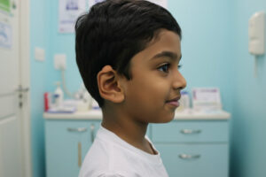
Imaging in otology plays a pivotal role in diagnosing and managing various ear disorders. Advanced imaging techniques, such as CT scans and MRIs, provide detailed visualizations of the ear’s intricate structures, enabling precise identification of abnormalities. Under the expert guidance of Dr. Shree Rao, these imaging modalities are utilized to assess conditions like chronic otitis media, cholesteatoma, and vestibular schwannomas. By leveraging these technologies, otologists can develop targeted treatment plans, enhancing patient outcomes and ensuring comprehensive ear health management.
Preoperative Assessment and Planning
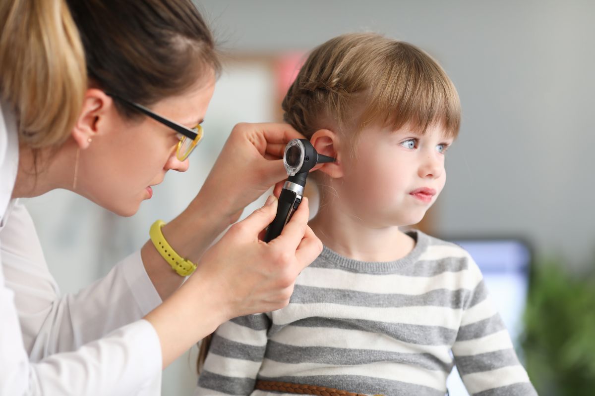
Preoperative assessment and planning play a crucial role in ensuring the success of ear surgeries. By employing advanced imaging techniques, otolaryngologists can thoroughly evaluate the patient’s ear anatomy, identify any abnormalities or pathology, and develop a precise surgical plan tailored to the individual’s needs.
During the preoperative assessment, the otolaryngologist conducts a comprehensive evaluation of the patient’s medical history, symptoms, and previous imaging studies, if available. This information helps guide the selection of appropriate imaging modalities and informs the surgical approach.
Imaging modalities commonly used in preoperative assessment include computed tomography (CT) scans, magnetic resonance imaging (MRI), and high-resolution CT (HRCT). CT scans provide detailed images of the bony structures of the ear, making them invaluable for assessing conditions such as chronic otitis media and temporal bone fractures. MRI, on the other hand, offers superior soft tissue contrast, making it ideal for visualizing soft tissue pathology like acoustic neuromas and identifying vascular abnormalities.
The information obtained from these imaging studies allows the otolaryngologist to accurately diagnose the patient’s condition, assess the extent of disease, and plan the surgical approach accordingly. For example, in cases of chronic otitis media with cholesteatoma, preoperative imaging helps identify the location and size of the cholesteatoma, determine the extent of bone erosion, and plan the appropriate surgical technique for complete removal.
Additionally, preoperative imaging plays a crucial role in identifying any anatomical variations or abnormalities that may impact surgical outcomes. For instance, in patients undergoing cochlear implantation, imaging helps assess cochlear morphology, identify cochlear anomalies, and determine electrode array placement, ensuring optimal implantation and postoperative outcomes.
Imaging Modalities in Ear Surgery
Computed Tomography (CT) Scan: CT scans are widely utilized in ear surgery for their ability to provide high-resolution images of the temporal bone and surrounding structures. They offer detailed visualization of bony anatomy, making them invaluable for assessing conditions such as chronic otitis media, temporal bone fractures, and congenital anomalies. CT scans help surgeons identify the extent of disease, evaluate bone erosion, and plan the surgical approach accordingly.
Magnetic Resonance Imaging (MRI): MRI is particularly useful for visualizing soft tissue structures within the ear, including the cochlea, vestibular apparatus, and neural elements. It offers superior soft tissue contrast and is highly effective in diagnosing conditions such as acoustic neuromas, vestibular schwannomas, and vascular abnormalities. MRI helps surgeons assess the involvement of soft tissue structures, plan surgical approaches, and monitor postoperative outcomes.
High-Resolution CT (HRCT): HRCT combines the benefits of CT scanning with enhanced spatial resolution, allowing for detailed visualization of delicate structures within the temporal bone. It is commonly used in complex cases requiring precise anatomical delineation, such as cochlear implantation, lateral skull base surgery, and stapes surgery. HRCT provides surgeons with detailed information about bony landmarks, anatomical variations, and disease extent, facilitating meticulous surgical planning and intraoperative navigation.
Cone Beam CT (CBCT): CBCT is a specialized imaging technique that offers volumetric imaging of the temporal bone with lower radiation exposure compared to conventional CT scans. It is particularly useful in minimally invasive ear surgeries, such as endoscopic ear surgery and percutaneous cochlear implantation. CBCT provides real-time imaging during surgery, enabling surgeons to confirm instrument placement, assess anatomical landmarks, and ensure accurate surgical navigation.
Intraoperative Imaging: Intraoperative imaging modalities, such as intraoperative CT and fluoroscopy, provide real-time visualization during ear surgeries, allowing surgeons to confirm surgical landmarks, assess instrument placement, and verify the completeness of surgical procedures. These modalities enhance surgical precision, reduce the risk of complications, and improve patient outcomes by ensuring optimal surgical execution.
Specific Surgical Applications
Tympanoplasty is a surgical procedure performed to repair a perforated tympanic membrane (eardrum). It involves grafting tissue onto the perforation site to restore the integrity of the eardrum, improve hearing, and prevent recurrent infections. Tympanoplasty is indicated for patients with chronic otitis media, traumatic perforations, or congenital defects affecting the tympanic membrane.
Mastoidectomy is a surgical procedure used to treat chronic mastoiditis, cholesteatoma, or other conditions involving the mastoid bone. It involves the removal of diseased mastoid air cells to eliminate infection, prevent complications such as mastoid abscesses or meningitis, and restore normal ear anatomy and function. Mastoidectomy may be performed as an open procedure or using minimally invasive techniques such as endoscopic mastoid surgery.
Stapedectomy is a surgical procedure performed to treat otosclerosis, a condition characterized by abnormal bone growth in the middle ear, leading to conductive hearing loss. During stapedectomy, the stapes bone is removed and replaced with a prosthesis to restore sound transmission to the inner ear. Stapedectomy is highly effective in improving hearing outcomes and is associated with low complication rates when performed by experienced surgeons.
Cochlear implantation is a surgical procedure indicated for patients with severe to profound sensorineural hearing loss who derive limited benefit from hearing aids. It involves implanting a device directly into the cochlea to bypass damaged hair cells and stimulate the auditory nerve, allowing for sound perception and speech comprehension. Cochlear implantation requires meticulous surgical technique and comprehensive postoperative rehabilitation to optimize outcomes.
Lateral skull base surgery encompasses a range of procedures aimed at treating tumors, vascular malformations, and other lesions affecting the temporal bone, jugular foramen, and cerebellopontine angle. These surgeries may involve approaches such as translabyrinthine, retrosigmoid, or middle fossa approaches, depending on the location and extent of pathology. Lateral skull base surgery requires a multidisciplinary approach involving neurotologists, neurosurgeons, and skull base surgeons to achieve optimal outcomes while preserving neurological function.
Ossiculoplasty is a surgical procedure used to repair or reconstruct the ossicular chain within the middle ear, which may be damaged or disrupted due to trauma, infection, or congenital anomalies. It involves replacing or repairing the ossicles (malleus, incus, and stapes) to restore sound conduction and improve hearing. Ossiculoplasty may be performed using autologous tissue grafts, synthetic prostheses, or biocompatible materials, depending on the patient’s specific needs and anatomy.
Postoperative Imaging
Assessment of Surgical Outcomes: Postoperative imaging allows for the detailed evaluation of surgical results. For instance, in tympanoplasty, imaging can confirm the proper placement and integration of the graft, ensuring that the tympanic membrane has been successfully repaired. In cochlear implantation, imaging verifies the accurate positioning of the electrode array within the cochlea.
Detection of Complications: Early identification of postoperative complications is essential for prompt management and optimal patient outcomes. Imaging modalities such as CT and MRI can detect issues such as residual cholesteatoma, infection, or fluid accumulation in the middle ear. These findings can guide timely medical or surgical interventions.
Monitoring Healing and Recovery: Postoperative imaging is useful in monitoring the healing process over time. For patients who have undergone mastoidectomy or lateral skull base surgery, serial imaging can track the resolution of inflammation, the regrowth of healthy tissue, and the stability of any implanted materials or prostheses.
Guiding Further Treatment: In some cases, postoperative imaging may reveal the need for additional procedures. For example, if ossiculoplasty outcomes are suboptimal, imaging can help identify ossicular discontinuity or prosthesis displacement, guiding revision surgery. Similarly, imaging can assist in planning further interventions for residual or recurrent pathology detected after initial surgery.
Conclusion - The Role of Imaging in Planning and Performing Ear Surgeries
Imaging in planning and performing ear surgeries is indispensable, offering unparalleled insights into the complex anatomy of the ear. With the expertise of Dr. Shree Rao, imaging techniques like CT and MRI scans facilitate accurate diagnosis and surgical precision, ultimately improving patient outcomes. By integrating advanced imaging into surgical planning, otologists can enhance the effectiveness and safety of ear surgeries, underscoring the critical role of imaging in modern otological practice.

Why consult EarSurgeon, Dr. Shree Rao?
Dr. Shree Cuddapah Rao is acclaimed as one of the best pediatric ENT specialists in Hyderabad. With 10+ years of deep domain experience in the field of ENT, she is the director at Dr. Rao’s ENT Super Specialty Hospital. She underwent specialized training in Rhinoplasty / Facial Plastic surgery at Singapore General Hospital, Singapore. She also underwent advanced training in cochlear implant surgery under Padmashri Dr. Milind V Kirtane and had a Fellowship in a cochlear implant. Having performed over 200 successful cochlear implants for patients worldwide, Dr. Shree Cuddapah Rao is also the recipient of several prestigious accolades in the domain of ENT. Dr. Shree Rao is one of the best ent doctor in hyderabad, to book an appointment click here.
Are you looking for
then you have landed at right place!